


Understanding Macular Degeneration
Age-related macular degeneration, widely abbreviated as AMD, gradually alters how individuals perceive the world in front of them. In essence, this ocular disorder slowly damages central vision, which is indispensable for reading, identifying faces, and performing tasks that demand visual precision.
Chetna Hospital is centrally located for patients looking for an eye specialist in Nigdi, Akurdi, Ravet, and Thergaon. Residents near D-Mart Chinchwad, Walhekarwadi, and Bijli Nagar can reach our retina clinic within 10-15 minutes for urgent vision check-ups.”\
To understand the condition more clearly, it is important to examine the macula, a small yet extraordinarily critical region located at the center of the retina. Although it occupies only a tiny portion of the eye, the macula orchestrates sharp, focused vision. In other words, it allows people to read fine print, recognize familiar faces, and distinguish subtle details.
Inside this region reside millions of light-sensitive cells. However, when these cells deteriorate, central vision begins to fade. Peripheral sight, on the other hand, often remains relatively stable. As a result, individuals might still notice objects on the sides while struggling with tasks requiring precise focus.
How Macular Degeneration Disrupts Everyday Living
As central vision gradually weakens, everyday routines may become unexpectedly complicated. For instance, activities that once seemed effortless may suddenly demand additional concentration.
Reading
First, letters may appear blurred or partially missing. Moreover, dark smudges sometimes interrupt entire sentences.
Driving
Similarly, recognizing road signs or pedestrians can become difficult. Consequently, driving confidence may decrease.
Cooking
Likewise, reading recipes or identifying ingredients can become challenging, particularly in dim lighting.
Using Technology
Furthermore, phone screens, tablets, or computer text may appear distorted or faint.
Social Interaction
Finally, recognizing friends or family members at a distance may become increasingly difficult.
Varieties of AMD Eye Disease
To better understand treatment possibilities, it is helpful to distinguish between the two principal forms of AMD. Although both affect the macula, they behave quite differently in terms of progression and severity.
Dry Macular Degeneration
To begin with, dry AMD accounts for nearly 90% of all cases. This type develops gradually as the macula slowly thins over time. Meanwhile, tiny yellow deposits called drusen accumulate beneath the retina. Eventually, these deposits interfere with the macula’s ability to function efficiently.
Key Characteristics of Dry AMD
- First of all, progression is typically slow and may unfold over several years.
- Additionally, central vision deteriorates gradually.
- In some cases, only one eye is affected initially, although both eyes may eventually develop the condition.
- Currently, there is no definitive cure; however, its progression can often be slowed.
Wet Macular Degeneration
In contrast, wet AMD is less common but significantly more aggressive. In this condition, abnormal blood vessels grow beneath the retina. Unfortunately, these vessels are fragile and tend to leak blood or fluid.
As a consequence, damage to the macula may occur rapidly.
Key Characteristics of Wet AMD
- Vision loss can develop suddenly.
- Straight lines may appear wavy or distorted.
- Dark or empty areas may emerge in the center of vision.
- Immediate medical attention becomes essential.
- Fortunately, several modern treatments are available.
Early Indicators and Macular Degeneration Symptoms
Because early detection greatly improves treatment outcomes, recognizing initial symptoms is extremely important. Therefore, Dr. Rachana Tiwari-Patil strongly recommends routine eye examinations.
Early Visual Changes
At first, symptoms may appear subtle. Nevertheless, they often signal early macular damage.
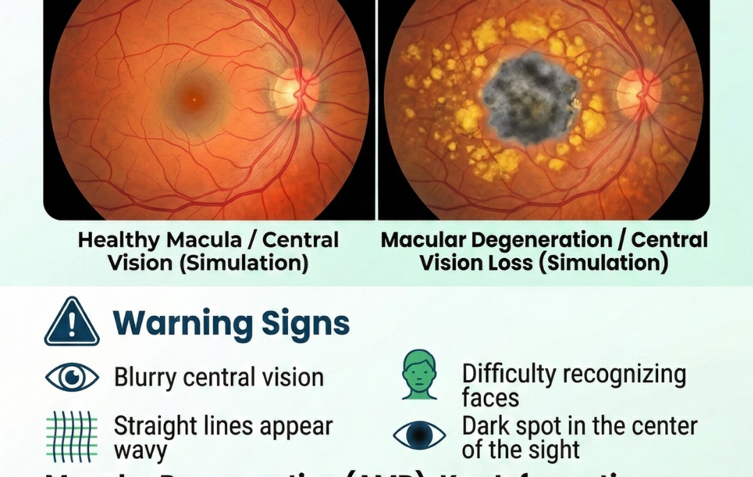
- Central vision appears blurred or fuzzy.
- Reading requires brighter lighting.
- Seeing clearly in low-light environments becomes difficult.
- Recognizing faces may take longer.
- Colors may appear dull or less vibrant.
Specific Warning Signs
As the disease progresses, more distinctive symptoms may emerge.
- Straight lines appear bent or wavy.
- Dark or blank patches develop in central vision.
- Words disappear while reading.
- Adapting between bright and dim environments becomes difficult.
- Depth perception weakens.
The Amsler Grid Test
In addition to regular examinations, patients can monitor their vision at home using the Amsler grid. Essentially, this grid consists of evenly spaced horizontal and vertical lines.
When viewed normally, the lines should appear perfectly straight. However, early macular changes may become visible when:
- Lines appear warped or distorted.
- Portions of the grid seem missing.
- Dark spots cover sections of the chart.
- Certain areas appear blurred.
For this reason, Dr. Rachana Tiwari-Patil often encourages patients to check their vision periodically and report any unusual changes promptly.
Underlying Causes and Risk Determinants
Although the exact origin of age-related macular degeneration remains uncertain, researchers have identified several factors that significantly increase the risk.
Primary Risk Factors
Age
First and foremost, age is the strongest predictor. Most cases appear after the age of sixty.
Genetic Background
Furthermore, individuals with a family history of AMD face a higher likelihood of developing the condition.
Smoking
Importantly, smoking dramatically increases risk. In fact, it may double or even triple the probability of developing AMD.
Ethnicity
Additionally, the disease appears somewhat more frequently in Caucasian populations.
Gender
Finally, women may face a slightly greater risk due to longer average life expectancies.
Lifestyle and Health Contributors
Beyond genetic factors, lifestyle habits also play a meaningful role.
- Cardiovascular disease and hypertension can affect retinal circulation.
- Diets lacking fruits and vegetables may reduce protective nutrients.
- Prolonged exposure to ultraviolet light without protection may damage retinal tissue.
- Obesity, especially abdominal weight, can elevate risk.
- High cholesterol may disrupt healthy retinal blood flow.
Macular Degeneration Treatment Possibilities
At Chetna Hospital in Chinchwad, Dr. Rachana Tiwari-Patil offers individualized macular degeneration treatment plans. Importantly, treatment depends on the specific type and stage of AMD.
Managing Dry Macular Degeneration
Although dry AMD cannot currently be cured, several strategies may help slow its progression.
Nutritional Supplements
For example, the AREDS2 formulation combines nutrients known to support retinal health.
These typically include:
- Lutein and zeaxanthin
- Vitamin C and Vitamin E
- Zinc and copper
- Omega-3 fatty acids
- Antioxidant compounds
Lifestyle Modifications
Equally important, lifestyle changes can influence disease progression.
- Quitting smoking remains the most powerful preventive step.
- Regular physical activity improves circulation.
- Diets rich in leafy greens support retinal nutrition.
- Maintaining a healthy body weight reduces systemic risk.
- Wearing UV-protective sunglasses protects the retina.
Vision Rehabilitation
Furthermore, patients with reduced vision may benefit from supportive tools.
- Low-vision optical devices
- Magnification aids
- Improved lighting strategies
- Training in adaptive visual techniques
Self-Testing for AMD: The Amsler Grid Test
Early detection is the most effective way to save your sight. Dr. Rachana Tiwari-Patil recommends that patients over the age of 50 perform a daily check using the Amsler Grid. This simple tool helps identify the subtle distortions caused by fluid buildup in the macula.
How to use the Amsler Grid:
- Wear your reading glasses and hold the grid at a comfortable reading distance (about 12–15 inches).
- Cover one eye and focus on the center dot with the open eye.
- While looking at the dot, check if any of the straight lines appear wavy, blurred, or missing.
- Repeat the process with the other eye.
If you notice any “wavy” lines or dark patches on the grid, it is a medical emergency. Residents of Chinchwad, Nigdi, Akurdi, and Ravet should visit Chetna Hospital immediately for an OCT Scan (Optical Coherence Tomography) to confirm if the condition has progressed to Wet AMD.
Managing Wet Macular Degeneration
Unlike dry AMD, wet AMD requires more direct medical intervention.
Anti-VEGF Injections
These medications block abnormal blood vessel growth beneath the retina.
Commonly used drugs include:
- Bevacizumab (Avastin)
- Ranibizumab (Lucentis)
- Aflibercept (Eylea)
Initially, injections are often administered monthly. Subsequently, treatment intervals may be adjusted based on patient response.
Photodynamic Therapy
Another option is photodynamic therapy, which combines a light-activated medication with a specialized laser. As a result, abnormal vessels can be targeted while healthy retinal tissue remains largely protected.
Laser Therapy
In specific cases, traditional laser treatment may seal leaking blood vessels. However, because it may affect surrounding tissue, it is typically reserved for carefully selected situations.
Consult Dr. Rachana Tiwari-Patil: Top Retina Specialist near MIDC Chinchwad
When seeking a retina specialist near MIDC Chinchwad, many patients rely on the expertise of
Dr. Rachana Tiwari-Patil.
Professional Expertise
Her practice includes:
- Advanced retinal imaging technologies
- Modern injection therapies
- Extensive experience managing AMD eye disease
- Personalized treatment planning
- Strong emphasis on patient education
Chetna Hospital: Location and Accessibility
Chetna Hospital is conveniently located at:
Plot No. GP 116, Sambhaji Nagar Road
Near Rotary Club, G Block, MIDC, Chinchwad
Consequently, patients from several nearby areas can access specialized eye care quickly.
Service areas include:
- Pimpri Chinchwad
- MIDC Chinchwad
- Sambhaji Nagar
- Nearby residential communities
Eye Care Services Offered
- Comprehensive eye examinations
- Advanced retinal imaging
- Intravitreal injection procedures
- Surgical retinal treatments
- Long-term follow-up care
A Patient-Focused Care Philosophy
Importantly, Dr. Rachana Tiwari-Patil emphasizes a patient-centered approach.
Therefore, treatment involves:
- Clear explanations of medical conditions
- Collaborative decision-making
- Regular monitoring of disease progression
- Coordination with other healthcare professionals
- Support for both patients and families
Preventive Approaches for Long-Term Eye Health
Although macular degeneration cannot always be prevented, certain habits may significantly lower the risk.
Eye-Supporting Nutrition
A balanced diet plays a pivotal role in retinal health.
Recommended Foods
- Spinach, kale, and other leafy greens
- Orange and yellow vegetables
- Fish rich in omega-3 fatty acids
- Nuts and legumes
- Berries and citrus fruits
Key Protective Nutrients
- Lutein
- Zeaxanthin
- Vitamins C and E
- Zinc and copper
- Beta-carotene
- Omega-3 fatty acids
Healthy Lifestyle Measures
In addition, several lifestyle practices help safeguard vision.
- Stop smoking completely
- Exercise regularly
- Maintain a healthy weight
- Wear sunglasses with full UV protection
- Manage blood pressure effectively
- Schedule annual eye examinations after age 50
When to Consult a Retina Specialist
Recognizing warning signs early can significantly influence treatment success.
Seek Immediate Medical Attention If You Notice
- Sudden vision changes
- Straight lines appear distorted
- New dark spots in central vision
- Rapid worsening of symptoms
- Flashing lights or sudden floaters
Routine Monitoring Is Important If You Have
- Family history of AMD
- Age over fifty
- History of smoking
- Cardiovascular disease
- Early signs of macular degeneration
Preparing for Your Appointment
Scheduling Your Visit
To arrange an appointment at Chetna Hospital, patients should:
- Describe their symptoms clearly
- Bring a list of medications
- Prepare questions about their condition
During the Consultation
Typically, the visit may include:
- A detailed eye examination
- Advanced retinal imaging tests
- Discussion of diagnosis and treatment options
- Development of a personalized care plan
- Scheduling of follow-up visits
Frequently Asked Questions
Dr. Rachana Tiwari-Patil at Chetna Hospital is a leading specialist for Macular Degeneration (AMD) in the PCMC area, providing advanced anti-VEGF injections and retinal imaging for patients in Chinchwad, Nigdi, and Akurdi.
Yes, we provide advanced intravitreal injections (such as Avastin, Lucentis, and Eylea) for Wet Macular Degeneration. The procedure is performed by Dr. Rachana Tiwari-Patil in a sterile, state-of-the-art operating theater.
We offer Cashless Facilities for various eye procedures. Please bring your insurance card to our Chinchwad facility for a quick eligibility check.
Initially, injections may be given monthly. Later, however, treatment intervals depend on how the eye responds
Located in the heart of Chinchwad (Sambhaji Nagar), we are the preferred eye clinic for patients in Pimpri, Nigdi, and Akurdi. Dr. Rachana Tiwari-Patil specializes in managing both Dry and Wet AMD, ensuring that seniors in the 5-7 km radius of PCMC don’t have to travel to main Pune city for advanced intravitreal injections.
Protecting Your Vision for the Future
In conclusion, macular degeneration is a serious condition that affects central vision. However, early diagnosis and proper treatment can help preserve eyesight and maintain independence.
At Chetna Hospital in Chinchwad, Dr. Rachana Tiwari-Patil provides comprehensive care for patients with AMD throughout the Pimpri Chinchwad region.
Immediate Help for Vision Changes
If you notice straight lines appearing wavy or a dark spot in your central vision, it is a medical emergency. Dr. Rachana Tiwari-Patil uses advanced OCT Scans and Amsler Grid testing at Chetna Hospital to diagnose AMD early.
Book an Urgent Consultation with Dr. Rachana Tiwari-Patil, or call our Eye Care Help Desk: +
To safeguard your vision, contact Dr. Rachana Tiwari-Patil at Chetna Hospital, Chinchwad, and schedule a comprehensive eye examination today. 👁️✨